Beyond Organ-on-a-Chip: Understanding Microphysiological Systems (MPS)
As biomedical research moves toward more human-relevant and animal-free testing methods, Microphysiological Systems have emerged as one of the most promising innovations in preclinical research.
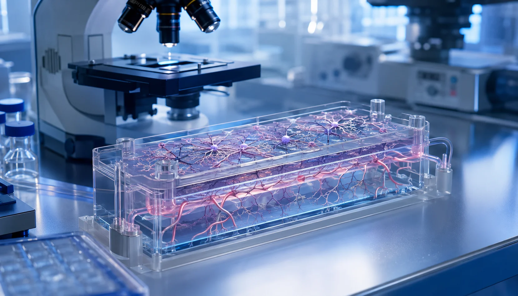
Designed to better replicate the structure and function of human tissues, Microphysiological Systems combine advances in cell biology, tissue engineering, and microfluidics to create models that more closely resemble human physiology than conventional in vitro methods.
Rather than replacing existing technologies, MPS integrate approaches such as organoids and organ-on-a-chip platforms into a broader framework for studying human biology and evaluating new therapies.
What Are Microphysiological Systems?
Microphysiological Systems are advanced laboratory models that recreate key biological functions of human tissues and organs outside the body. Unlike traditional two-dimensional cell cultures, MPS provide a controlled environment where cells can interact in three dimensions and respond to mechanical and biochemical cues that more closely resemble those found in vivo.
The term Microphysiological Systems is commonly used as an umbrella concept that includes organ-on-a-chip technologies, organoids, and other engineered tissue models.
These technologies share a common goal: generating more predictive and human-relevant data for drug discovery, disease modeling, toxicology studies, and therapeutic development.
How Are MPS Different from Organ-on-a-Chip?
Although the terms Microphysiological Systems and organ-on-a-chip are sometimes used interchangeably, they do not describe exactly the same concept.
| Feature | Microphysiological Systems | Organ-on-a-Chip |
|---|---|---|
| Definition | A broad category of advanced models designed to recreate aspects of human tissue and organ physiology. | A microfluidic device developed to reproduce the function of a particular organ or tissue. |
| Technology Scope | Can include organoids, engineered tissues, organ-on-a-chip platforms, and interconnected multi-tissue systems. | Primarily focuses on microfluidic platforms containing living cells and controlled fluid flow. |
| Research Use | Supports studies involving single tissues, multiple organs, systemic interactions, and complex disease mechanisms. | Often used to investigate the behavior and response of a specific organ or tissue. |
Organ-on-a-chip platforms are therefore considered one important type of MPS. This broader perspective enables researchers to select the most appropriate model for the biological question being studied, whether the project involves a single organ, multiple interacting tissues, or a complex disease pathway.
Why Are Microphysiological Systems Important?
One of the most significant challenges in drug development is predicting how a therapy will behave in humans. Conventional cell cultures often oversimplify biological processes, while animal models may not accurately reproduce human physiology or patient-specific responses.
Microphysiological Systems help address these limitations by creating models that better mimic tissue architecture, cellular interactions, biochemical signaling, and physiological conditions.
More Predictive Data
Human-relevant tissue models can help researchers evaluate how potential therapies may behave before progressing to clinical studies.
Earlier Safety Assessment
Potential toxicity and adverse biological responses can be investigated during earlier stages of preclinical development.
Reduced Animal Reliance
MPS contribute to the development of alternative research strategies that support the reduction and refinement of animal testing.
By producing more physiologically relevant information, MPS can improve confidence in preclinical findings and support better-informed decisions before clinical trials.
Applications of MPS in Biomedical Research
Microphysiological Systems are now being used across a growing range of biomedical research areas. Their adaptable structure allows researchers to design models around specific tissues, diseases, compounds, and biological questions.
Drug Discovery
MPS can support the evaluation of new compounds by providing more physiologically relevant information about efficacy, safety, and tissue response.
Disease Modeling
Human-derived cells can be used to study complex disease mechanisms and observe how biological changes develop within controlled laboratory environments.
Toxicology Studies
Advanced tissue models can improve the assessment of human responses to pharmaceuticals, chemicals, and other test substances.
As MPS technologies continue to evolve, they are also becoming valuable tools for precision medicine by helping researchers develop and evaluate therapies that better reflect individual patient biology.
Looking Ahead
Microphysiological Systems are reshaping the future of biomedical research by helping bridge the gap between conventional cell culture and human physiology. As advances in microfluidics, stem cell biology, tissue engineering, and biomaterials continue, these models are expected to play an increasingly important role in the development of safer medicines and more informative preclinical studies.
Their continued development may also support more ethical research practices by expanding the availability of scientifically robust alternatives to traditional testing methods.
Conclusion
Microphysiological Systems represent a new generation of human-relevant research models. By integrating technologies such as organ-on-a-chip platforms, organoids, and advanced engineered tissues, MPS provide researchers with more predictive tools for drug development, disease research, and toxicology.
As the scientific community continues to adopt innovative alternatives to conventional testing methods, Microphysiological Systems are positioned to become a cornerstone of modern biomedical research.
Advancing Human-Relevant Biomedical Research
OmicaCell supports the development of advanced cellular models designed to help researchers explore human biology, disease mechanisms, and therapeutic responses in more relevant laboratory environments.
Patient-Derived Organoids: Bringing Personalized Medicine into the Laboratory
Patient-derived organoids offer researchers a powerful way to examine disease biology and potential treatment responses using three-dimensional models created from an individual patient’s own cells.
Every patient is unique, and so is their response to treatment. This variability has driven growing interest in personalized medicine, where therapies are tailored to an individual’s biology rather than relying entirely on a one-size-fits-all approach.
One of the most promising tools supporting this transition is the patient-derived organoid. Created from a patient’s own cells, these three-dimensional models can closely resemble the structure and function of the original tissue, allowing researchers to study diseases and evaluate potential therapies in a more clinically relevant way.
What Are Patient-Derived Organoids?
Patient-derived organoids are miniature, three-dimensional tissue models grown from cells obtained through biopsies or surgical samples. Unlike conventional two-dimensional cell cultures, PDOs preserve many of the biological characteristics, genetic profiles, and cellular diversity found in the original tissue.
Because patient-derived organoids maintain patient-specific characteristics, they can provide researchers with models that more accurately represent human biology, individual disease features, and clinically relevant treatment responses.
These characteristics make PDOs valuable tools for biomedical research, disease modeling, drug development, and the continued advancement of personalized medicine.